Mom has this fast-growing, dome-shaped pink bump with a hard plug in the center on her forearm. The clinic is closed for the weekend. What is this?
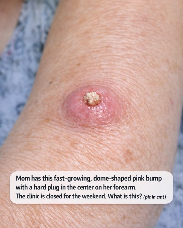
If you suddenly notice a fast-growing, pink, dome-shaped bump with a firm-looking plug in the middle, I completely understand why that would send you into a weekend spiral. I’m the person who can stay calm through a packed workweek, but let one odd skin spot show up on a Friday night and I’m Googling with one hand while reheating leftovers with the other. A bump like this on the forearm can look dramatic, especially when it seems to pop up quickly over 2 to 8 weeks, and one possible cause doctors think about is something called a keratoacanthoma.
The tricky part is that keratoacanthoma can resemble other skin growths, including squamous cell carcinoma, so this is not really a “watch it forever and hope for the best” situation. In this article, I’ll walk through what keratoacanthoma usually looks like, why it tends to worry clinicians, what you can do safely over the weekend, what not to do, and which red flags mean you should skip waiting for the clinic to reopen and get urgent care sooner.
1. What keratoacanthoma usually looks like
Keratoacanthoma is a skin growth that often appears as a round, raised, dome-shaped bump that is pink, red-pink, or skin-colored. A classic clue is the center: it often has a firm, rough, crusty, or horn-like keratin plug. Many are about 5 to 20 millimeters across when first noticed, but some can grow to 1 to 2 centimeters or more in a fairly short time.
One reason people notice it so quickly is the growth speed. Unlike a spot that has quietly sat there for 2 years, keratoacanthoma often enlarges over a matter of weeks. A person may say, “This was barely there a month ago, and now it looks like a tiny volcano.” That fast change is exactly why it gets attention.
2. Why the forearm is a common place for it
Keratoacanthomas often show up on sun-exposed skin. That includes the forearms, backs of the hands, lower legs, face, and sometimes the ears. The forearm is a very believable location because it gets years of incidental sunlight driving to work, gardening, walking the dog, or just sitting near a window.

In my part of the Midwest, a lot of us underestimate sun exposure because we think only beach days “count.” But daily exposure adds up. Ten to 20 minutes here, another 15 minutes there over years, especially without SPF 30 or higher, can contribute to cumulative skin damage.
3. Why it can’t be diagnosed for sure from appearance alone
This is the part I would not downplay: keratoacanthoma can look very similar to squamous cell carcinoma, a type of skin cancer. In fact, many clinicians treat a suspected keratoacanthoma as something that needs prompt in-person evaluation because distinguishing the two may require a biopsy or complete removal.
Even if a bump seems to fit the “dome with a central plug” pattern perfectly, photos and descriptions are not enough to confirm it. Other possibilities can include a wart-like growth, an inflamed cyst, a cutaneous horn over another lesion, or other less common tumors. The exact diagnosis depends on the exam and often the pathology report.
4. The typical age and risk pattern
Keratoacanthoma is more common in adults, especially middle-aged and older adults, and more often in people with a history of significant sun exposure. It may also be more likely in people with fair skin, those who burn easily, and people whose immune systems are suppressed by medicines or medical conditions.
There are also cases linked with prior skin injury, though sun damage is the big pattern. If your mom has had a lot of outdoor time, prior precancers, or previous skin cancers, that raises the importance of having it checked quickly. If she has never had anything like this before, it still deserves evaluation because the appearance and rapid growth matter more than the skin history alone.
5. The “fast-growing” part is one of the biggest clues
A lot of harmless skin spots grow very slowly, sometimes over years. Keratoacanthoma is different because it can enlarge rapidly over 2 to 6 weeks, sometimes a little longer. People will often describe it as “suddenly there” even though technically it built up over days to weeks.
If you have measurements, write them down. For example, “It was about 4 millimeters two weeks ago and now it’s 11 millimeters.” That timeline is very useful for the doctor. If you do not have exact measurements, compare it to a pencil eraser, pea, or blueberry, but a ruler photo taken from a normal distance is even better.
6. What you should do while the clinic is closed
If it is not bleeding heavily, not causing severe pain, and your mom otherwise feels fine, the safest weekend plan is usually simple: leave it alone, protect it, and arrange an appointment as soon as the clinic reopens. If possible, call first thing Monday morning and mention that it is a rapidly growing lesion with a central keratin plug, because those words may help staff understand why it should be seen sooner rather than later.
If the spot rubs on sleeves or furniture, cover it loosely with a clean, nonstick bandage. A small non-adherent pad and paper tape or a gentle self-adhesive wrap can help. Change the dressing once daily or sooner if it gets wet or dirty. Wash the area gently with mild soap and lukewarm water, then pat dry. No scrubbing.
7. What not to do this weekend
Do not squeeze it, pick at the center plug, cut it, freeze it at home, burn it with any gadget, or apply acid-based wart remover. Those products often contain salicylic acid or similar ingredients and can badly irritate the skin, distort the lesion, and make the doctor’s evaluation more difficult.
I know it is tempting to “just get the hard center out,” especially if it looks like a plug that should pop free. But traumatizing it can trigger bleeding, pain, infection, and a less clear exam. The central material is usually keratin, not something that should be dug out at the kitchen counter under bright lights at 10 p.m.